Ocular surface

Dry eye disease — getting your comfort back

Dry eye disease affects several million people in France and accounts for a large share of eye care visits. It shows up as stinging, burning, a foreign-body sensation or paradoxical watering. Far from being a mere nuisance, it is a chronic inflammation of the tear film and the ocular surface that requires appropriate care. At the practice, I offer a comprehensive diagnosis and a personalized, step-by-step treatment plan, ranging from simple measures to innovative treatments such as IPL or topical cyclosporine.

OSDI questionnaire In-office diagnosis Step-by-step treatment IPL & cyclosporine
Illustration evoking the feeling of sand in the eyes — a hallmark symptom of dry eye disease
Understanding

What is dry eye disease?

Dry eye is much more than a nuisance: it is an imbalance of the tear film. According to the TFOS DEWS III report (2025, the most recent international reference), it is a multifactorial, symptomatic disease characterized by a loss of homeostasis of the tear film and/or ocular surface, in which tear film instability, hyperosmolarity, inflammation and neurosensory abnormalities are the etiological factors.

The tear film: three essential layers

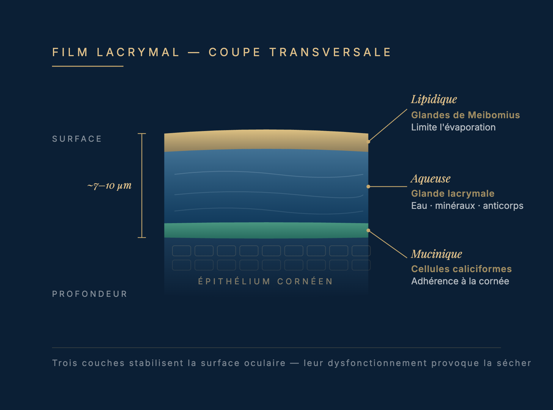
Your tears are not just a drop of water. They form a film made up of three layers:

  • Lipid layer — meibomian glands. Limits evaporation.
  • Aqueous layer — lacrimal gland. Provides water, minerals and antibodies.
  • Mucin layer — goblet cells. Helps the film adhere to the cornea.
Cross-sectional diagram of the tear film: lipid layer (meibomian glands, ~surface), aqueous layer (lacrimal gland, water · minerals · antibodies), mucin layer (goblet cells, adhesion to the cornea), total thickness ~7-10 µm.

A dysfunction in any one of these layers is enough to cause dry eye. In most cases of dry eye (about 50 to 85% depending on the study), the lipid layer (meibomian glands) is affected — this is the main cause of evaporative dry eye (TFOS DEWS II/III reports).

The two main mechanisms

Hyposecretion — not enough tears (Sjögren's, age, medications). Evaporation — meibomian gland dysfunction (MGD), the leading cause.

  • Meibomian gland dysfunction: secretions that are too thick or blocked
  • Rapid loss of the lipid film
  • Hyperosmolar tears = inflammation
  • Paradox: reflex watering without lubrication

Diagnostic key: Tear hyperosmolarity (> 308 mOsm/L) triggers inflammation, which in turn worsens dryness. It is a vicious cycle.

Recognizing dry eye

Symptoms & triggers

Typical sensations
  • Stinging, itching
  • Burning sensation
  • Gritty, sandy or foreign-body sensation
  • Paradoxical watering
  • Fluctuating vision (better after blinking)
  • Sensitivity to light
  • Eyelids stuck together in the morning
Triggering factors
  • Prolonged screen work
  • Reduced blinking
  • Dry, air-conditioned or heated environments
  • Wind, drafts
  • Close-up reading
  • End of the day
  • Contact lens wear
  • Makeup on the eyelid margin
Who is at risk?

Key risk factors

Demographic & hormonal

  • Age > 50 — very common after 60
  • Female sex — 2-3× more common
  • Menopause — drop in estrogen
  • Hormone therapy
  • Pregnancy — temporary adaptation

Systemic diseases

  • Sjögren's syndrome (Gougerot-Sjögren disease)
  • Lupus, rheumatoid arthritis
  • Rosacea
  • Diabetes
  • Graft-versus-host disease (GVHD)

Environment & medications

  • Screens
  • Contact lenses
  • Antihistamines, antidepressants
  • Beta-blockers, anticholinergics
  • Isotretinoin (acne)
  • Eye drops with preservatives (benzalkonium chloride)

Lifestyle

  • Smoking
  • Chronic UV exposure without protection
  • Dry air, air conditioning
At the practice

A comprehensive dry eye diagnosis

I use several tools to pinpoint the cause and severity of your dry eye, and tailor the treatment accordingly.

OSDI questionnaire & medical history
We go over your symptoms, when they started and what triggers them. The OSDI is a validated score of perceived severity, used as a baseline to assess your response to treatment.
Slit lamp — Eyelid margin & meibomian glands
I look for inflammation, debris and the quality of meibomian gland secretions. Normal vs. thickened secretions = diagnosis of the mechanism.
TBUT — Tear Break-Up Time (short = abnormal)
After instilling fluorescein, I time how long it takes for the tear film to break up. This measures tear film instability, a sign of lipid layer dysfunction.
Fluorescein staining (Oxford scale)
This dye highlights areas of damaged epithelium. It helps assess severity and points toward the cause.
Stepwise treatment

Step-by-step treatment (DEWS III)

I offer a tailored, stepwise treatment based on the TFOS DEWS III 2025 management algorithm (Jones, Craig et al., Am J Ophthalmol). We start simple, then step up the treatment according to your profile and clinical response.

Step 1 — Lifestyle & Education
20-20-20 rule (every 20 min, look into the distance for 20 sec). Conscious blinking. Eyelid hygiene. Omega-3 2-3 g/day. Hydration. Well-positioned screen. Avoid direct air conditioning.
Step 2 — Artificial tears & simple topical care
PRESERVATIVE-FREE tears — a must (hyaluronic acid, HP-guar, trehalose, lipid-based). 3-6×/day. Gels/ointments at night. Daily eyelid care (warm compresses, massage, hygiene).
Step 3 — Cyclosporine & anti-inflammatory drugs
Steroidal or non-steroidal anti-inflammatory drugs. Cyclosporine (several strengths available from hospital and retail pharmacies) — once or twice a day. Oral doxycycline.
Step 4 — Specialized treatments
Intense pulsed light (IPL) therapy. Dissolvable or permanent punctal plugs (reduce tear drainage). Autologous serum in severe cases. Therapeutic scleral lenses. Indications tailored to your profile and your response to the previous steps.

Strategy: I start at the step matching your OSDI severity. If the response is insufficient after 4-6 weeks, I move up to the next step. Be patient with timelines: cyclosporine brings its first benefits after 1 to 2 months.

Dr HAGE's research

Dryness, eye rubbing, a vicious cycle

During my time as Assistant Spécialiste at the Hôpital National des Quinze-Vingts (Paris) under Prof. Christophe Baudouin, I devoted a large part of my research to the still widely overlooked link between dry eye, itching and eye rubbing. My publications shed light on a key mechanism behind the chronic course of ocular surface diseases.

First author · 2023

From ocular itching to eye rubbing : a review of the literature

Journal Français d'Ophtalmologie · 2023;46(2):173-184

A comprehensive review of ocular itching and its link with eye rubbing: initial dry eye symptoms, onset of scratching, release of inflammatory cytokines, corneal damage, and perpetuation of the itch. A novel parallel with dermatological models helps explain why treating dry eye also means protecting the cornea.

DOI ↗ PubMed ↗
First author · 2023

EYERUBBICS — The Eye Rubbing Cycle Study

Journal of Clinical Medicine · 2023;12(4):1529

Our prospective study of 153 patients highlights, for the first time, the cognitive and addictive behavioral dimension of eye rubbing in keratoconus and ocular surface diseases. Article featured by Ophthalmology Times, which supports a stronger educational approach with my patients.

DOI ↗ PubMed ↗
All my scientific publications ↗
Innovative treatment

IPL treatment for meibomian gland dysfunction

IPL (Intense Pulsed Light) is an established treatment option for meibomian gland dysfunction (MGD), with a level of evidence now supported by several case series and reviews (Toyos, Reinstein, Cote/Cochrane). To learn all about this technology, read my article “IPL for dry eyes: the complete guide”.

How does it work?

Intense light pulses are absorbed by inflamed blood vessels and glands:

  • Reduces vascular inflammation
  • Liquefies meibomian gland secretions
  • Reduces the Demodex population
  • Restores the microcirculation

Effectiveness: improvement in symptoms and tear parameters (TBUT, OSDI) is reported in most patients after 3-4 sessions (Toyos 2015, Reinstein 2018).

Protocol

  • Sessions: 3-4, spaced 1 to 4 weeks apart
  • Duration: 15-20 min / both eyes
  • Anesthesia: None (mild discomfort possible)
  • Precautions: Avoid sun for 24 hours. Normal activities afterward.
  • Results: Gradual. Peak at 1 month. Annual maintenance.
Full IPL treatment page ↗
Special case

Dry eye after LASIK & refractive surgery

All corneal refractive surgery causes temporary corneal denervation, which can worsen dry eye in the first few months. The vast majority of these symptoms are mild and temporary; they resolve with proper care. Here is what I offer my refractive surgery candidates.

~50%
Mild, temporary symptoms in the first month
3 to 6 months
Usual time for corneal nerve recovery
< 5%
Symptoms persisting at 1 year with careful pre-operative screening

Why this temporary dryness?

The surgery cuts or alters some of the corneal nerve endings. This temporarily reduces the nerve feedback that stimulates tear secretion and reflex blinking.

These nerves gradually regenerate, over 3 to 6 months on average. SMILE and PRK spare corneal innervation more than LASIK and carry a lower risk of postoperative dry eye — these are the techniques I favor for patients prone to dry eye.

Source: TFOS DEWS III (2025) — Iatrogenic Dry Eye chapter.

How I anticipate and treat it

  • Before surgery — full ocular surface assessment (TBUT, examination of the eyelids and ocular surface, OSDI). If subclinical dry eye is present, I treat it 4 to 6 weeks before the operation.
  • Choice of technique — SMILE or PRK rather than LASIK for at-risk profiles.
  • Day 0 to Day 7 — preservative-free tears 6-8×/day, visual rest, emphasis on blinking.
  • Month 1 to Month 3 — close follow-up, adjustment of drop frequency.
  • If symptoms persist — cyclosporine 0.1%, punctal plugs, IPL if associated MGD.

With this strategy, more than 95% of my patients regain lasting comfort at 12 months.

Pre-refractive surgery assessment ↗ See my refractive surgery techniques
Validated questionnaire

Assess your dry eye (OSDI-6)

The OSDI-6 is the short version of the Ocular Surface Disease Index, recommended by the TFOS DEWS III (2025) report. Six quick questions help assess the severity of your dry eye and guide your care.

Over the past few weeks, how often have you experienced…

Q1. Sensitivity to light?
Q2. A gritty or foreign-body sensation?
Q3. Eye pain or discomfort?
Q4. Blurred or fluctuating vision?
Q5. Discomfort when reading?
Q6. Eyes irritated by wind or drafts?
Your care pathway

Managing dry eye

1
Consultation & Diagnosis
History, OSDI, full examination (slit lamp, TBUT, staining, eyelids). Identification of the cause and severity.
2
Treatment plan
Tailored recommendations. Start at step 1-2 (hygiene, artificial tears). Explanation of timelines and realistic expectations.
3
Follow-up & Adjustments
Reassessment at 2-4 weeks. If the response is insufficient, move to the next step (cyclosporine, IPL). Patience with timelines.
4
Stabilization & Maintenance
Comfort restored, better quality of life. Annual follow-up. You play an active role in keeping your comfort.
Frequently asked questions

FAQ — Dry eye disease

Next step

Are your eyes bothering you every day?

A comprehensive ocular surface assessment allows me to identify the mechanism behind your dry eye and offer a tailored, lasting, personalized treatment.

Cabinet OPHTALIFE — Boulogne-Billancourt (next to Paris)
Former assistant surgeon Hôpital des Quinze-Vingts (Paris)
Book online via Doctolib
Learn more

Related pages on the ocular surface

Bibliography

Scientific references

This page is based on international guidelines. All sources can be verified.

  1. 1

    Wolffsohn JS, Benítez-Del-Castillo JM, Loya-Garcia D, et al. TFOS DEWS III: Diagnostic Methodology. Am J Ophthalmol. 2025;279:387-450.

  2. 2

    Jones L, Craig JP, Markoulli M, et al. TFOS DEWS III: Management and Therapy. Am J Ophthalmol. 2025;279:289-386.

  3. 3

    Stapleton F, Argüeso P, Asbell P, et al. TFOS DEWS III: Digest. Am J Ophthalmol. 2025;279:451-553.

  4. 4

    Perez VL, Chen W, Craig JP, et al. TFOS DEWS III: Executive Summary. Am J Ophthalmol. 2025;282:135-145.

  5. 5

    Schiffman RM, Christianson MD, Jacobsen G, et al. Reliability and validity of the Ocular Surface Disease Index (OSDI). Arch Ophthalmol. 2000;118(5):615-621.

  6. 6

    Cote S, Ahuja M, Chen Z, et al. Intense pulsed light (IPL) therapy for meibomian gland dysfunction. Cochrane Database Syst Rev. 2020;3(3):CD013559.

  7. 7

    Toyos R, McGill W, Briscoe D. Intense pulsed light treatment for dry eye due to meibomian gland dysfunction. Photomed Laser Surg. 2015;33(1):41-46.

  8. 8

    Craig JP, Chen YH, Turnbull PR. Prospective trial of IPL for meibomian gland dysfunction. Invest Ophthalmol Vis Sci. 2015;56(3):1965-1970.

  9. 9

    Hage A, Knoeri J, Leveziel L, et al. From ocular itching to eye rubbing: a review of the literature. J Fr Ophtalmol. 2023;46(2):173-184.

  10. 10

    Hage A, Knoeri J, Leveziel L, et al. EYERUBBICS: The Eye Rubbing Cycle Study. J Clin Med. 2023;12(4):1529.

The content of this website is for information purposes only and does not replace a medical consultation.